Create an access opening to reach the lesion.
Using etching as the foundation, add selective enamel removal when necessary to access the surface barrier sufficiently for resin infiltration.
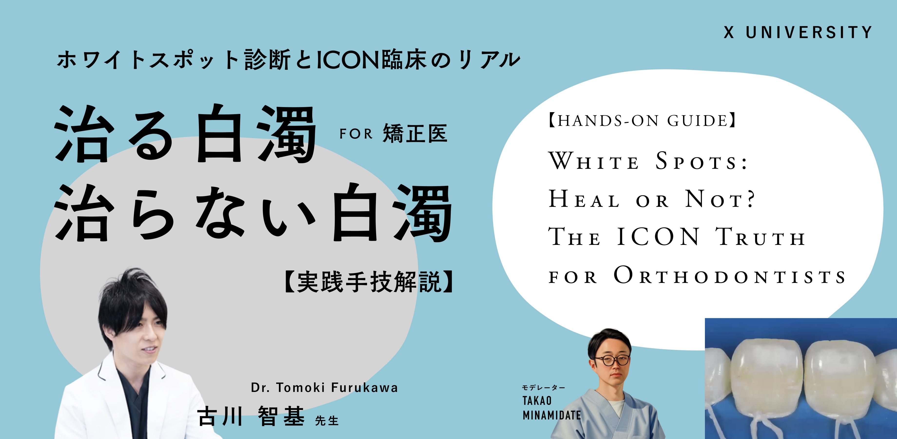
X UNIVERSITY / ENGLISH ARCHIVE EDITION
Dr. Tomoki Furukawa / Moderator: Dr. Takao Minamidate
ARCHIVE ONLY · NO LIVE ACCESS · LIVE SESSION IN JAPANESE
Learn how to distinguish white spots that may respond to resin infiltration from lesions that require another approach. The course moves beyond the product instructions into diagnosis, lesion depth, surface access, clinical checkpoints, and recovery decisions when treatment does not go as planned.
FILM 03 / DIAGNOSIS
Not every white lesion is the same. Dr. Furukawa begins with the diagnostic distinction between demineralized and non-demineralized lesions before ICON is considered.
The real question
White spot lesions are not a single diagnosis. Etiology, depth, and surface condition differ. If you simply follow the steps without recognizing these differences, you may not achieve the expected change.
Is this white opacity likely to improve with ICON?
How do you differentiate demineralized white spot lesions from fluorosis and MIH?
What do you assess from the appearance after applying ethanol?
If whiteness persists, how do you reassess and what do you choose next?
What is ICON?
ICON is a micro‑invasive treatment that infiltrates a low‑viscosity resin into porous enamel. The name comes from “Infiltration CONcept.” It is not a method of simply painting the tooth white, nor does it involve removing all opaque enamel and restoring those areas.
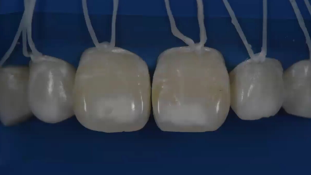
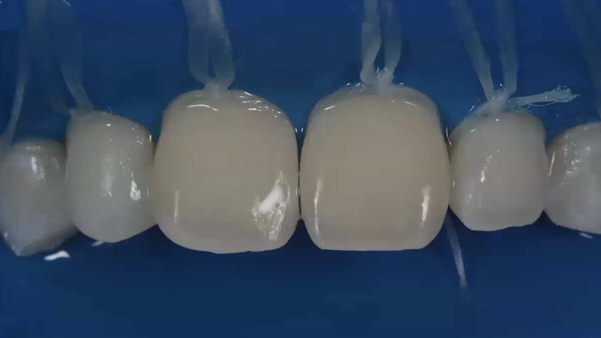
Using etching as the foundation, add selective enamel removal when necessary to access the surface barrier sufficiently for resin infiltration.
Observe changes on wetting with Icon Dry. If the white opacity does not improve adequately, do not proceed and re-evaluate access.
Low‑viscosity resin is infiltrated into the lesion, then light‑cured, re‑applied and polished. Moisture control and assessment at each step influence the outcome.
ICON is a product name.
Diagnosis and interim evaluation strongly influence the outcome.
Lesions that look similarly white may differ in depth and surface barrier depending on whether they are associated with demineralization, fluorosis, MIH, developmental disturbance, or trauma. ICON Smooth Surface is indicated for non-cavitated carious lesions and non-cavitated lesions associated with demineralization or mild-to-moderate fluorosis.
Not all white opacities improve the same way. Which lesions to select, how far to open the surface barrier, and which changes to confirm before proceeding—learning that clinical judgment is the focus of this seminar.
Sources: DMG Icon Smooth Surface product information, Instructions for Use, and Bourouni S, et al. Clin Oral Investig. 2021;25:4711-4719. doi:10.1007/s00784-021-03931-7. Diagrams were created independently by X UNIVERSITY from these sources. Follow the current instructions for use and regulatory requirements applicable in your jurisdiction.
Lecture preview
How much do post‑debonding opacities change over time? Using natural‑history data, we consider patient explanation and the need for intervention.
From pre-session planning we selected five clinical topics that serve as entry points to the lecture. Select a number to switch the video.
What you will learn
We clarify an approach to distinguish the underlying causes of white spots — demineralizing lesions, non‑demineralizing conditions such as fluorosis, MIH, and others.
Learn the meaning and limitations of clinically useful assessments such as the Wet-Dry Test, cross-polarized photography, and transillumination.
Observation, prevention, whitening, microabrasion, resin infiltration, restoration—connect these options into a single decision pathway.
Moisture control, surface treatment, etching, drying, infiltration — review the clinical observation points that are not obvious from the order in the instructions.
Which findings during evaluation justify proceeding? What changes should prompt re‑treatment? We define the checkpoints that influence success.
When opacity persists, appears banded, or a morphological defect develops, we will consider reassessment and alternative treatments, including failure cases.
For orthodontists
Orthodontic treatment does not end
when the teeth are aligned.
What patients see in the mirror is the final result of their orthodontic treatment.
After multi-bracket treatment—especially in cases with difficult plaque control—post-debonding white opacities can affect the final result. Whether the opacities arose during treatment or were pre-existing, the orthodontist must provide explanation and make clinical judgments.
Should you observe, continue preventive measures, start with whitening, choose ICON, or restore? Having a decision framework changes post‑treatment confidence and the quality of the outcome.
Recorded seminar
The recording walks through real clinical images to assess pathology, lesion depth, and indication.
Unfavorable outcomes are reviewed to identify missed checkpoints and recovery options.
Dr. Minamidate frames the questions from an orthodontist's perspective and discusses them with Dr. Furukawa.
Included archive resources
To translate the lecture into clinical practice, two take-home resources guide you from white-spot assessment to treatment selection.
Condense etiology, depth, surface status, and appearance into one framework to guide decisions from observation and prevention to infiltration and restoration.
From moisture control through assessment, infiltration, and finishing: make 'what to look for' at each step into a form you can verify chairside.
Those who complete the post-viewing survey also receive a patient information sheet and consent confirmation form. Downloadable clinical resources are currently provided in Japanese.
Speaker
Medical Corporation Taishi | Director, Wako City Dental Office
A reproducible protocol,
not a display of dexterity.
Dr. Furukawa is a comprehensive clinician whose practice spans general dentistry, esthetic restorative dentistry, implant dentistry, and orthodontics. This course is not a showcase of polished outcomes alone: it is designed to show orthodontists what to observe, how to sequence each step, and when to stop and reassess.
After graduating from Kyushu Dental University, he completed clinical training in the Department of General Dentistry at Aichi Gakuin University Hospital. He later served as vice director at SCDC Shizuoka Dental Clinic and as supervising dentist and vice director at Mizuho-dai Station Dental & Orthodontic Clinic before taking his current role. He is also involved in practice management and the education of younger clinicians.
Recommended for
Event details
14 SEP 202621:00-23:30 JST
Price & registration
FILM 14 / PROCEDURE
Insufficient preparation prevents the resin from reaching the lesion, while excessive preparation can leave a dark area. The key is deciding how much enamel to remove and when to proceed.
English-subtitled archive
US$50 USD
Archive-only access · No live access
Scheduled within 2–3 days after the Japanese live session
Register for the English archiveYou will continue to Stripe's secure checkout. Access instructions will be sent to your checkout email address.
Use the email address where you want to receive archive access.
A confirmation email explains the release schedule and access method.
The English-subtitled archive is scheduled within 2–3 days after the event.
Access remains active for 1 month from the English archive release date.
FAQ
Yes. It covers the material basics, case selection, technique, outcome assessment, and troubleshooting, with orthodontists who are less familiar with restorative procedures in mind.
Yes. It is also relevant to general dentists who provide orthodontic care or are interested in esthetic restorative treatment and white spot management.
Dental hygienists and other team members involved in prevention, monitoring, and patient communication may also find it useful. Diagnosis and treatment procedures remain the responsibility of a dentist.
It is scheduled for release within 2–3 days after the Japanese live seminar. Your 1-month viewing period begins on the archive release date.
No. This is an archive-only ticket and does not include live access. The live seminar is delivered in Japanese. The English-subtitled archive is scheduled for release within 2–3 days after the event and remains available for 1 month from release.
Participants receive the White Spot Decision Map and ICON Chairside Checklist. Those who complete the post-viewing survey also receive a patient information sheet and consent confirmation form. These downloadable clinical resources are currently provided in Japanese.
Yes. You can submit a clinical question during checkout. It may be addressed during the Japanese live seminar, but live access is not included with this ticket.
For paid registrations, use the receipt button in your purchase-confirmation email. If you cannot find it, enter your checkout email on the receipt delivery page; the link is sent only to that address. No payment receipt is issued for a zero-cost invitation.
Refunds are generally not available after purchase. Duplicate charges, payment errors, or an inability on our part to provide the archive will be reviewed individually.
Mastering White Spot & ICON
English-subtitled archive · US$50 · 1-month access from release
View archive registration